Sketch And Label Of A Cross Section Of A Long Bone : Schematic Diagram Of Long Bone Cross Section 47 Download Scientific Diagram. Label the parts of a long bone. An osteon is the basic functional and structural unit of mature compact bone. Two types of bone tissues in cross section of a long bone : 12 november 2008, 17:16 (utc) derivative work: Create a drawing of the bone section in your laboratory journal and label the areas listed above.
This is the long central shaft. This is covered by a membrane of connective tissue called the periosteum.beneath the cortical bone layer is a layer of spongy cancellous bone.inside this is the medullary cavity which has an inner core of bone marrow, it contains nutrients and help in formation of cells, made up of yellow marrow in. 12 november 2008, 17:16 (utc) derivative work: A = epiphysis b = diaphysis c = articular cartilage d = periosteum f = compact bone g = medullary cavity (yellow marrow) h = endosteum j = epiphyseal line (growth plate) coloring worksheet for this image. Lamellar bone makes up the compact or cortical bone in the skeleton, such as the long bones of the legs and arms.
Bones And Muscles Theschoolrun from i.ytimg.com The structure of a long bone allows for the best visualization of all of the parts of a bone (figure 6.7). Related posts of cross section of a long bone bone test anatomy and physiology. A long bone has a shaft and 2 ends. As shown in figure 2. Forms the larger rounded ends of long bones. 1) from a mechanical standpoint, bone is historically the most studied tissue, and 2) due to 1) and the simpler behavior of bone compared to soft tissues, more is known about bone mechanics in relation to its structure. Osteons are oriented parallel to the diaphysis of the long bone. The original can be viewed here:
The structure of a long bone consists of several sections:.
Use colored pencils to draw and label the following structures as they appear using the 40x objective, or by looking at an image from the internet. Smartdraw includes 1000s of professional healthcare and anatomy chart templates that you can modify and make your own. Use snap chat and take/label a picture. This is for two reasons: (do not copy and paste a picture from the text or internet.) The ends of a long bone contain spongy bone and an epiphyseal line. Label the parts of a long bone. Click on the tags below to find other quizzes on the same subject. The walls of the diaphysis are composed of dense and hard compact bone. A long bone has two parts: A long bone illustrates both types of bone. Then, fill in the table below to describe each. The diaphysis and the epiphysis.
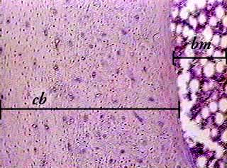
Label the parts of a long bone. A long bone has two parts: Label the haversian canal, osteocyte (mature bone cell) in lacuna, and canaliculi. This image shows compact bone in cross section. Label the membrane that lines the cavity and the membrane that covers the outside surface.
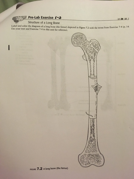
Solved Pre Lab Exercise 5 Structure Of A Long Bone Label Chegg Com from media.cheggcdn.com 12 november 2008, 17:16 (utc) derivative work: Make sure learners follow all the criteria for a biological drawing. Continue to label this drawing as you explore the inside of the bone. The diaphysis is the tubular shaft that runs between the proximal and distal ends of the bone. Marks should be deducted for shading or colouring. Draw and label a longitudinal section of a long bone. Use snap chat and take/label a picture. Once we stop growing (between 18.
This image shows compact bone in cross section.
The hollow region in the diaphysis is called the medullary cavity, which is filled with yellow marrow. Smartdraw includes 1000s of professional healthcare and anatomy chart templates that you can modify and make your own. 1) from a mechanical standpoint, bone is historically the most studied tissue, and 2) due to 1) and the simpler behavior of bone compared to soft tissues, more is known about bone mechanics in relation to its structure. 12 november 2008, 17:16 (utc) derivative work: Use snap chat and take/label a picture. The outer shell of the long bone is made of cortical bone also known as compact bone. As shown in figure 2. External circumferential lamellae, osteon, central canal, perforating canals, lacuna, canaliculi, concentric lamellae. The walls of the diaphysis are composed of dense and hard compact bone. A long bone has a shaft and 2 ends. Related posts of cross section of human bone diagram human back muscles and bones. The ends of a long bone contain spongy bone and an epiphyseal line. Sketch and label a cross section of a bone.
Draw and label a longitudinal section of a long bone. Sketch a longitudinal section through a long bone and label the following structures de epiphysim ercavi periosteum, co p pseen, compact bune.no red bone marrow, and yellow bone marrow he provides a epiphysis riedullary activity 4: Bone test anatomy and physiology 12 photos of the bone test anatomy and physiology anatomy and physiology bone lab test, anatomy and physiology bone markings test, anatomy and physiology bone practical test, anatomy and physiology bone tissue test, anatomy and physiology test on bone tissue, bone, anatomy and. This image shows compact bone in cross section. 1) from a mechanical standpoint, bone is historically the most studied tissue, and 2) due to 1) and the simpler behavior of bone compared to soft tissues, more is known about bone mechanics in relation to its structure.
Bone Compact Decalcified C S from www.austincc.edu Bone matrix and cells bone matrix osseous tissue is a connective tissue and like all connective tissues contains relatively few cells and large amounts of extracellular matrix. The hollow region in the diaphysis is called the medullary cavity, which is filled. A long bone has two parts: A long bone is a bone that has greater length than width. Some, mostly older, compact bone is remodelled to form these haversian systems (or osteons).the osteocytes sit in their lacunae in concentric rings around a central haversian canal (which runs longitudinally).the osteocytes are arranged in concentric rings of bone matrix called lamellae (little plates), and their processes run in interconnecting canaliculi. The walls of the diaphysis are composed of dense and hard compact bone. The head of each end of a long bone consists largely of spongy bone and is covered with hyaline cartilage. As shown in figure 2.
A long bone has two parts:
Forms the larger rounded ends of long bones. A cross section of a human long bone. A long bone has two parts: Make a pencil sketch and use markers or colored pencils to add details. The walls of the diaphysis are composed of dense and hard compact bone. Plates of cartilage, also known as growth plates which allow the long bones to grow during childhood. Label the parts of a long bone. Label the membrane that lines the cavity and the membrane that covers the outside surface. Sketch a longitudinal section through a long bone and label the following structures de epiphysim ercavi periosteum, co p pseen, compact bune.no red bone marrow, and yellow bone marrow he provides a epiphysis riedullary activity 4: External circumferential lamellae, osteon, central canal, perforating canals, lacuna, canaliculi, concentric lamellae. The original can be viewed here: Learners should accurately draw a long bone, resembling that in figure 6.24. The diaphysis and the epiphysis.
Share :
Post a Comment
for "Sketch And Label Of A Cross Section Of A Long Bone : Schematic Diagram Of Long Bone Cross Section 47 Download Scientific Diagram"

Post a Comment for "Sketch And Label Of A Cross Section Of A Long Bone : Schematic Diagram Of Long Bone Cross Section 47 Download Scientific Diagram"